
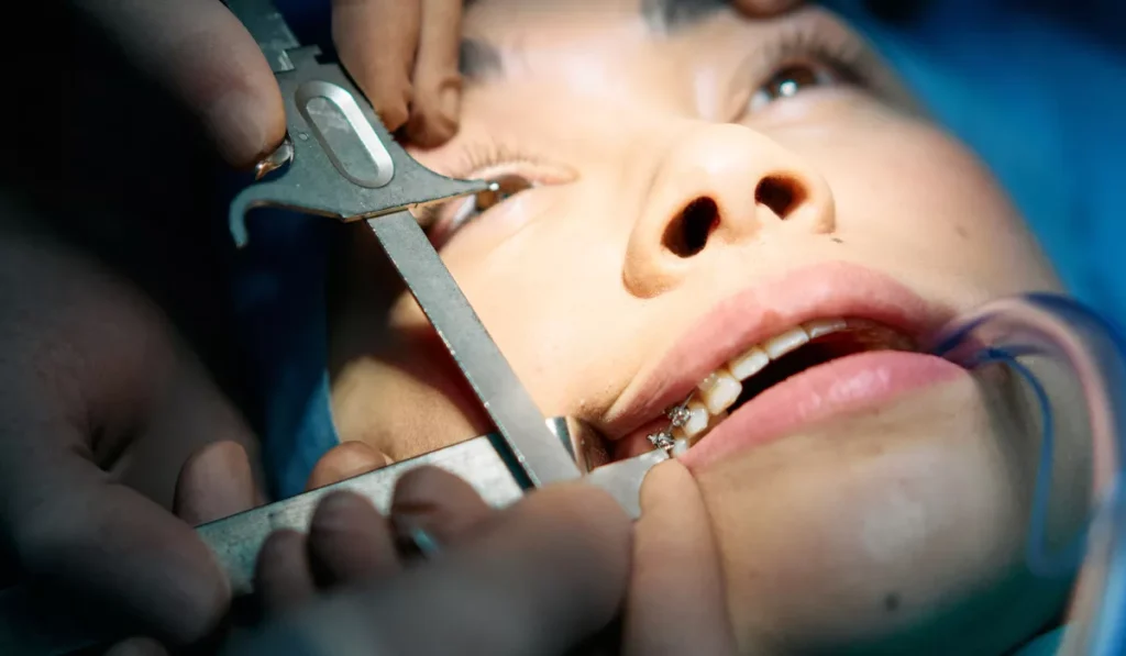
For more than 20 years, Brent Chapman lived with a form of blindness that half a lifetime of surgery couldn’t fix. This year, doctors in British Columbia solved it using something almost nobody would think to look at: one of his own teeth.
Chapman, now 34, underwent one of the first tooth-in-eye surgeries ever performed in Canada, a procedure so rare that only a few hundred people worldwide have ever received it.
A Childhood Reaction That Changed Everything
Chapman’s story began at age 13, when he suffered a rare and severe autoimmune reaction after taking a standard dose of ibuprofen. The condition, known as Stevens-Johnson syndrome, causes the immune system to attack the body’s own skin and mucous membranes, triggering injuries similar to severe burns across the body, including the surface of the eyes.
One of those injuries became infected and destroyed his left cornea, the eye’s transparent outer dome, entirely. Corneal damage also cost him most of the vision in his right eye.
Over the following two decades, Chapman underwent more than 50 surgeries, including 10 separate attempts at corneal implants in his right eye. Most failed. Some temporarily restored partial vision, but none permanently fixed the underlying damage.
A Procedure That’s Existed Since the 1960s, Just Never in Canada
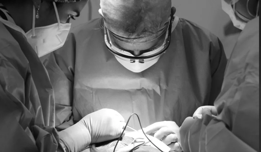
Doctors eventually proposed osteo-odonto-keratoprosthesis, more simply known as tooth-in-eye surgery, to restore vision in Chapman’s right eye. The technique itself isn’t new, it’s been performed since the 1960s, but Chapman’s case marked the first time it had ever been carried out in Canada.
The procedure works by removing one of the patient’s own teeth and implanting it into the eye socket, where it acts as a stable platform for a small, transparent plastic lens. That lens effectively replaces the damaged cornea, allowing light to reach the eye again.
Good candidates for the surgery typically have a damaged cornea but a healthy retina and optic nerve, the light-sensitive structures at the back of the eye still need to function properly for the procedure to restore usable vision. According to a 2022 study following 59 patients who underwent the surgery, 94% retained their vision long-term, based on follow-up data spanning as long as 30 years after the operation.
How the Surgery Actually Worked
Dr. Greg Moloney, a clinical associate professor of ophthalmology at the University of British Columbia, performed the first stage of the procedure on Chapman and two other Canadian patients in February 2025.
Moloney began by removing one of Chapman’s canine teeth, along with a thin layer of surrounding bone, carefully preserving its blood and oxygen supply. The tooth was then shaved down into a small block, drilled through, and fitted with a plastic cylinder designed to hold the eventual lens. This sculpted tooth was implanted into Chapman’s cheek, where it remained for several months while soft tissue grew around it.
In June, surgeons removed the tooth from his cheek and implanted it directly into his right eye. Because the tooth and its surrounding tissue came from Chapman’s own body, the risk of rejection was significantly lower than with a synthetic implant.
From Blurred Movement to 20/30 Vision
Almost immediately after the eye surgery, Chapman could perceive movement for the first time in years. His vision sharpened gradually over the following month, though some visual distortion required an additional corrective surgery in August to adjust the lens placement.
By late August, tests with corrective glasses showed Chapman had achieved 20/30 vision, meaning objects a person with perfect eyesight could see clearly from 30 feet away, Chapman could see just as clearly from 20 feet.
Why This Case Stands Out

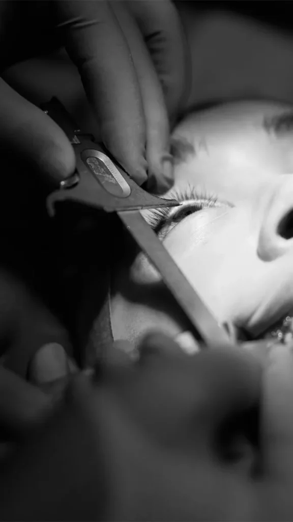
Tooth-in-eye surgery is considered a treatment of last resort for corneal blindness, reserved for cases where more conventional corneal transplants have already failed.
The full procedure spans more than 12 hours of surgical time, not counting the months required for tissue to grow around the implanted tooth in the patient’s cheek, and only a handful of specialists worldwide are trained to perform it.
The surgery has previously been performed in Australia, Chile, Egypt, Germany, India, Italy, Japan, Spain, the United Kingdom, and the United States.
To date, only a few hundred people globally have undergone the procedure, making Chapman’s case, and Canada’s first successful attempt, a notable addition to a very short list.
Frequently Asked Questions
What is tooth-in-eye surgery?
Tooth-in-eye surgery, medically known as osteo-odonto-keratoprosthesis (OOKP), is a rare procedure that uses a patient’s own tooth to support an artificial cornea. Surgeons attach a small plastic optical lens to the tooth before implanting it into the eye, allowing light to reach the retina and restore vision in carefully selected patients.
Who is a candidate for tooth-in-eye surgery?
The procedure is generally reserved for people with severe corneal blindness caused by injury, infection, or autoimmune diseases such as Stevens-Johnson syndrome. Candidates must still have a healthy retina and optic nerve, and tooth-in-eye surgery is usually considered only after conventional corneal transplants have failed.
Why do surgeons use a tooth instead of a synthetic implant?
A patient’s own tooth is far less likely to be rejected because it comes from their own body. It also provides a strong, long-lasting foundation for the artificial cornea, making it one of the most durable solutions available for certain types of severe corneal blindness.
How long does tooth-in-eye surgery take?
The procedure is completed in two stages over several months. During the first operation, surgeons prepare the tooth and implant it in the patient’s cheek, where living tissue grows around it. Months later, the reconstructed tooth is removed and implanted into the eye. Combined, the surgeries take more than 12 hours, not including the healing period between procedures.
How successful is tooth-in-eye surgery?
Although it is considered a last-resort treatment, tooth-in-eye surgery has produced impressive long-term results. A 2022 study following 59 patients found that 94% retained useful vision during follow-up periods of up to 30 years after surgery.
#wowfact |
Sources
- Live Science – Diagnostic Dilemma: Doctors restore a man’s vision by removing his tooth and implanting it in his eye
- International Journal / research references on osteo-odonto-keratoprosthesis (OOKP)